The brain consists of about 20 billion neurons which all generate electrical impulses. When these neurons work together in synchrony, tiny rhythmic, electrical potentials occur in the synapses which are specialised junctions between the neurons. The more neurons that work in synchrony, the larger the potential (amplitude) of the electrical oscillations measured in microvolts. The faster the neurons work together, the higher the frequency of the oscillations measured in Hertz. These two parameters: amplitude and frequency are the primary characteristics of brain waves.
These weak electrical signals can be measured by electrodes placed on the scalp using some conductive paste. After amplification by an EEG-amplifier, the signals are fed to a computer and analysed for amplitude and frequency. This is called electroencephalography (EEG).

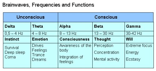
Brainwaves may be divided into 5 categories depending on the frequency: Delta waves (0.5-4 Hz) are dominant during coma and deep sleep. Theta waves (4-8 Hz) are associated with drives, emotions, trance states, and dream sleep. Alpha waves (8-13 Hz) reflect the brain’s idle state and are found in most people in the awake condition with closed eyes. Alpha waves are the prime indicators of conscious attention, and they represent the gate between the outer and the inner world and between the conscious and the unconscious. Beta waves (13-30 Hz) indicate an aroused, mentally alert and concentrated state. Finally, the fast Gamma frequencies (30-42 Hz) correlate with will, high energy states and ecstasy. Thus, both Delta and Theta waves reflect unconscious states, whereas Alpha and Beta waves indicate awake, conscious states. Finally recent research point to Gamma waves as the brain’s signature of higher states of consciousness.

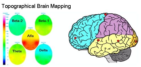
The Brainmaps are based on recordings of 8 channels of EEG. The following electrode placements are used according to the international 10-20 system: Fp1, Fp2, T3, T4, T5, T6, O1, O2, and Cz. The raw EEG is filtered through a band pass filter from 2-36Hz. In order to obtain baselines, the EEG is initially recorded during rest with closed and open eyes. After that the EEG is recorded during the activity or condition we want to study. After manual editing of the records for artifacts (removal of signals caused by muscle activity and eye movements) the computer performs an FFT frequency analysis usually of 60 seconds of artifact free record from each of the recorded conditions. Based on these analyses brainmaps showing the distribution of Delta, Theta, Alpha, Beta1, and Beta2 frequencies are constructed.
A brainmap gives a quick overview of the functional state of the brain. The brainmap above shows five coloured ovals displaying the distribution in the brain of five types of brain waves. The oval in the upper left corner actually shows a combination of Beta2 and Gamma frequencies. Each oval shows the brain seen from above, and the little tip at the top indicates the location of the nose. Each of the five ovals only displays one type of brain wave, however, all five types of waves are measured at the same time and at the same locations on the brain. Thus, the brainmap splits up the complex brain wave activity on five different displays in order to give a better overview.
The vertical, coloured scale to the left shows the power of the brain waves. Blue indicates low power, green and yellow medium, and red high power of the brain waves. The red colour on the middle oval, for example, indicates that Alpha are the most dominating waves in this brainmap, and are most predominant in the posterior (rear) part of the brain.
Usually activation of the brain will show up in the brainmap as decreased Theta and Alpha and increased Beta1 and Beta2 activity. On the other hand, if the subject relaxes and connects to his/her unconscious, we would tend to see an increase of Alpha and Theta activity.
Source: newbrainnewworld


0 comments:
Post a Comment